BIOPAD™ is a primary dressing composed of 100% native equine Type I collagen that can accelerate the closure of hard-to-heal wounds.
- Type I collagen of equine origin that keeps its native structure.1
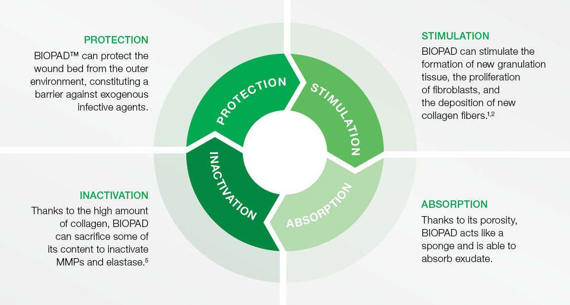
- Protects the wound bed from the outer environment, constituting a barrier against exogenous infective agents.
- Stimulates the formation of new granulation tissue, the proliferation of fibroblasts, and the deposition of new collagen fibers.1,2
- Absorbs wound exudate and can control minor bleeding.
References
- Rangaraj, A. & Harding, K & Leaper, D. (2011). Role of collagen in wound management. Wounds. 7.
- Karr, J & Taddei, A & Picchietti, S & Gambellini, G & Fausto, A & Giorgi, F. (2011). A Morphological and Biochemical Analysis Comparative Study of the Collagen Products Biopad, Promogram, Puracol, and Colactive. Advances in skin & wound care. 24. 208-16. 10.1097/01.ASW.0000397897.18003.ce.
- Beghé, F & Menicagli, C & Neggiani, P & Zampieri, A & Trallori, L & Teta, E & Rosini, S. (1992). Lyophilized non-denatured type-I collagen (Condress) extracted from bovine Achilles’ tendon and suitable for clinical use. International journal of tissue reactions. 14 Suppl. 11-9.
- Niebauer, G & Oz, M & Goldschmidt, M & Lemole, G. (1989). Simultaneous Use of Microfibrillar Collagen Hemostat and Blood Saving Devices in a Canine Kidney Perfusion Model. The Annals of thoracic surgery. 48. 523-7. 10.1016/S0003-4975(10)66854-3.
- Fleck, C & Chakravarthy, D. (2007). Understanding the Mechanisms of Collagen Dressings. Advances in skin & wound care. 20. 256-9. 10.1097/01.ASW.0000269310.00145.e2.
- Laghezza Masci, V & Taddei, A.R. & Gambellini, G & Giorgi, F & Fausto, A.M. (2017): Interaction And Cell Proliferation In Bioactive Collagen Matrices. Poster Session - SAWC Spring 2017
Orderinformation
BIOPAD Collagen dressing
sterile, individually-sealed
| Size | Item No. | HCPCS Code | Shipping Units (per box/case) |
|---|---|---|---|
| 5 x 5 cm (2 x 2 in.) | 153355 | A6021 | 3/168 |
| 10 x 10 cm (4 x 4 in.) | 153356 | A6021 | 1/34 |
Fields of application
- pressure ulcers
- donor sites and other bleeding surfaces
- dehisced surgical incisions
- draining wounds
- lacerations
- venous stasis ulcer
- diabetic ulcers
- partial- and full-thickness wounds
- post-laser surgery
- podiatric wounds
- surgical and traumatic wounds